Home

Opération possible Déchiqueté machine culs de sacs pleuraux album Héritier Ver de terre

Interprétation d'une radiographie thoracique en urgence – 2 – la plèvre |  thoracotomie
Interprétation d'une radiographie thoracique en urgence – 2 – la plèvre | thoracotomie

UE12 ED 2 : Radiographie thoracique : Les grands syndromes

Plevre
Plevre

Syndrome pleural
Syndrome pleural

Untitled
Untitled

Plevre
Plevre

POUMONS ET PLEVRES LES POUMONS

Plevre
Plevre

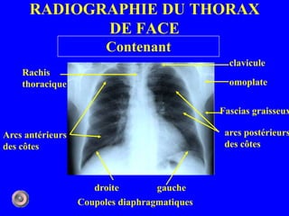
Radio du thorax - BIG PICTURE

Saga du poumon 11/. Le cul de sac pleural normal – Echo-Urgences

How to Interpret a Chest X-Ray (Lesson 6 - Diaphragm and Pleura) - YouTube

TROUBLES VENTILATOIRES RESTRICTIFS et RÉÉDUCATION KINÉSITHÉRAPEUTIQUE

cul de sac pleural D émoussé -opacité para-médiastinale D "en voile latine"  : thymus normal pour l'âge -hyperex
cul de sac pleural D émoussé -opacité para-médiastinale D "en voile latine" : thymus normal pour l'âge -hyperex
Pneumothorax spontane

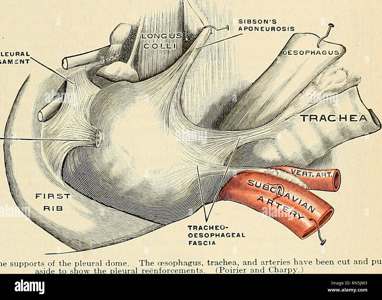
Anatomy, descriptive and applied. Anatomy. THi: PLEURJE 1183 and  Intercostal muscles is the costal pleura (pleura costalis); that which  covers the convex surface of the Diaphragm is the diaphragmatic pleura  [pleura
Anatomy, descriptive and applied. Anatomy. THi: PLEURJE 1183 and Intercostal muscles is the costal pleura (pleura costalis); that which covers the convex surface of the Diaphragm is the diaphragmatic pleura [pleura

POUMONS ET PLEVRES LES POUMONS

Pneumopéritoine visualisé à la face antérieure du foie chez le même... |  Download Scientific Diagram
Pneumopéritoine visualisé à la face antérieure du foie chez le même... | Download Scientific Diagram

Pr BOUKABACHE L Laboratoire d'Anatomie Humaine CHU Constantine

Interprétation d'une radiographie thoracique en urgence – 2 – la plèvre |  thoracotomie
Interprétation d'une radiographie thoracique en urgence – 2 – la plèvre | thoracotomie

Poumons et plèvres
Poumons et plèvres

1 anatomie et semiologie rdiologique (version 3)

cul de sac pleural D émoussé -opacité para-médiastinale D "en voile latine"  : thymus normal pour l'âge -hyperex
cul de sac pleural D émoussé -opacité para-médiastinale D "en voile latine" : thymus normal pour l'âge -hyperex

clichés thoraciques + interprétation +++ ( partie 1 ) - YouTube

Exploration radiologique du poumon - Medicinus

Interprétation d'une radiographie thoracique en urgence – 2 – la plèvre |  thoracotomie
Interprétation d'une radiographie thoracique en urgence – 2 – la plèvre | thoracotomie